
MRI-Compatiable Bioreactor for Cartilage Loading
Project Overview
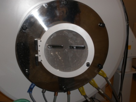
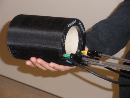
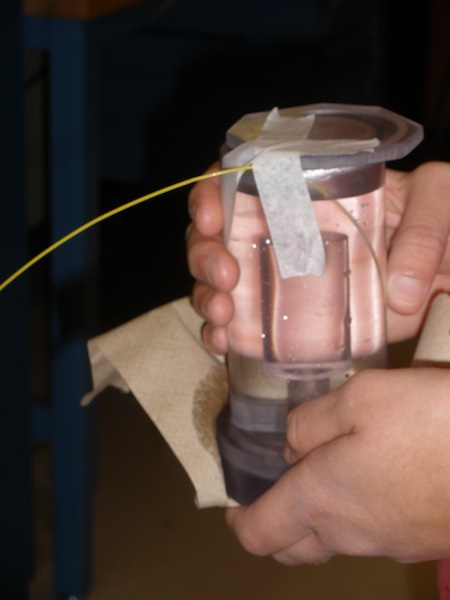
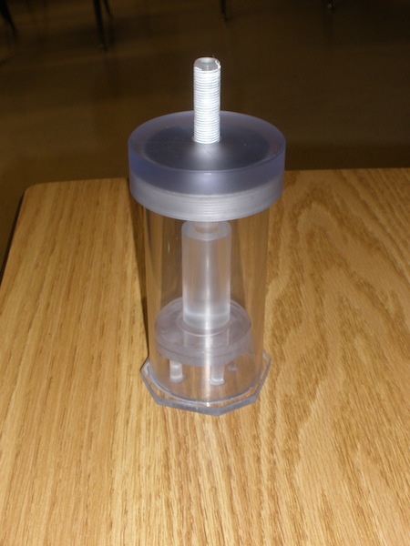
The objective of this project is to develop a bioreactor to secure engineered cartilage tissue during magnetic resonance (MR) scanning and provide mechanical loading during this scanning. Cartilage tissue cultures are currently grown in vitro for subsequent implantation into the body where the cartilage has been damaged by osteoarthritis or injury. Our bioreactor will be used to determine if the tissue is fully developed and mechanically stable. Unlike current testing, this bioreactor will be non-destructive to the tissue which will enable researchers to track the full development of a each cartilage sample.
Team Picture
Images
Files
- Midpoint Presentation (October 15, 2009)
- Mid-Semester Paper (October 25, 2009)
- PDS (December 5, 2009)
- Final Poster (December 5, 2009)
- Final Report (December 10, 2009)
Contact Information
Team Members
- Sarah Springborn - Team Leader
- Luisa Meyer - Communicator
- Beomkang Huh - BSAC
- Sarah Czaplewski - BWIG
Advisor and Client
- Prof. Walter Block - Advisor
- Prof. Wan-Ju Li - Client