
Computerized or mechanical 3D model for neuro-endoscopic teaching and practice
Project Overview
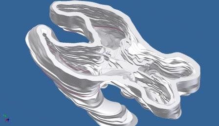
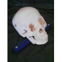
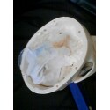
Our client, Dr. Bermans Iskandar, of the Department of Neurological Surgery at the UW Hospital, performs pediatric neurosurgeries. Currently, his medical students do not have a surgical simulator to practice endoscopic third ventriculoscopy to remove blockages in the cerebral aqueduct. A model of the ventricular system to teach and practice surgeries is necessary so that patients are not subject to initial trial surgeries by medical students. The model needs to be anatomically correct and allow the surgeon to practice control of their fine motor skills. The model should include insertion of the endoscope into the ventricles and be durable enough to train multiple students on the surgical techniques.
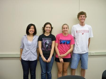
Team Picture
Images

Files
- Final Report (December 8, 2010)
- Final Poster (December 8, 2010)
- MidSemester Report (October 18, 2010)
- Progress Report Week 6 (October 19, 2010)
- Progress Report Week 5 (October 19, 2010)
- Progress Report Week 4 (October 19, 2010)
- Progress Report Week 3 (October 19, 2010)
- Progress Report Week 2 (October 19, 2010)
- Progress Report Week 1 (October 19, 2010)
- MidSemester Presentation PDF (October 19, 2010)
- Progress Report Week 7 (October 21, 2010)
- Progress Report Week 8 (October 28, 2010)
- PDS (November 4, 2010)
- Progress Report Week 9 (November 4, 2010)
- Progress Report Week 11 (November 19, 2010)
- Link to CAD drawings (December 7, 2010)
- Progress Report Week 12 (December 7, 2010)
Contact Information
Team Members
- Kimberli Carlson - Team Leader
- Courtney Krueger - Communicator
- Anyi Wang - BSAC
- Alan Meyer - BWIG
Advisor and Client
- Mitchell Tyler - Advisor
- Dr. Bermans Iskandar - Client
Related Projects
- Spring 2011: Computerized or mechanical 3D model for neuro-endoscopic teaching and practice
- Fall 2010: Computerized or mechanical 3D model for neuro-endoscopic teaching and practice