
Digital beam attenuator
Project Overview
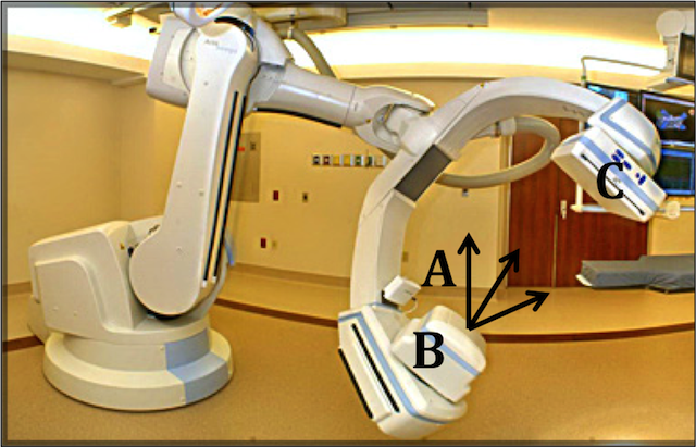
Conventional X-Ray Computed Tomography, a medical imaging procedure, employs a system that creates a three dimensional image of a patient by taking two dimensional X-Rays upon a revolution of the patient. This construction is used in various medical diagnoses. A current drawback to this procedure is the use of a uniform incident beam despite the fact that the transmission through the patient varies significantly from point to point. This results in a non-uniform signal-to-noise ratio, a sub-optimal distribution of x-ray scatter, and significantly higher dose than necessary to some regions and insufficient dose to others.
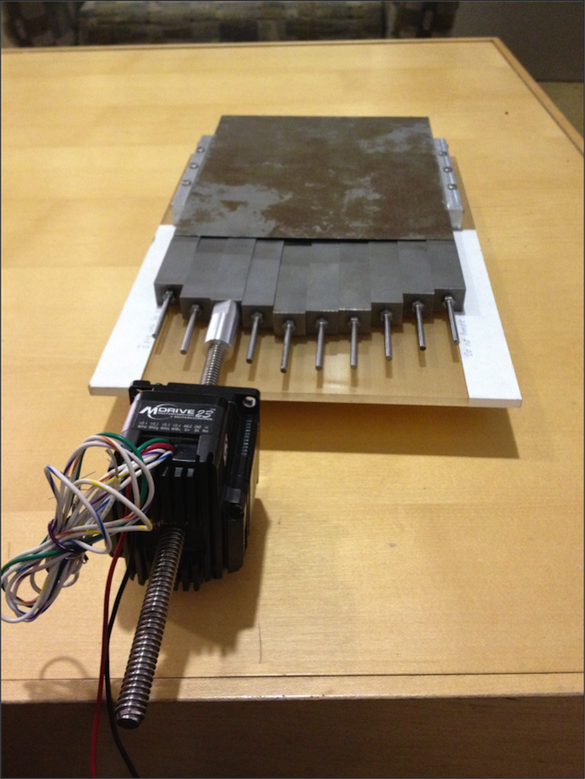
The primary aim of this project is to design, develop, and implement a series of attenuating wedges between the X-Ray focal point and the patient that continually attenuate the incident beams in order to compensate for the variance in the patient's body. Our team seeks to engineer and test the series of wedges so that they are able to independently adjust to the correct positions based on the data received from the computer (collected from a preliminary scan for each plane in the revolution) and continually attenuate the beam for an optimal scan.
Team Picture
Images
Files
- Poster (December 8, 2011)
- Final Paper (December 14, 2011)
- Test Plan (December 14, 2011)
- Midsemester Presentation (October 20, 2011)
- Midsemester Report (October 26, 2011)
Contact Information
Team Members
- Katherine Lake - Team Leader
- Alexander Eaton - Communicator
- Yue Hu - BSAC
- Sarvesh Periyasamy - BWIG
Advisor and Client
- Prof. Chris Brace - Advisor
- Prof. Chuck Mistretta - Client
Related Projects
- Spring 2012: Digital beam attenuator
- Fall 2011: Digital beam attenuator