
Radial expanding uterine cervical dilator

Project Overview

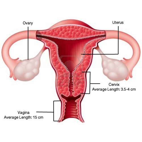
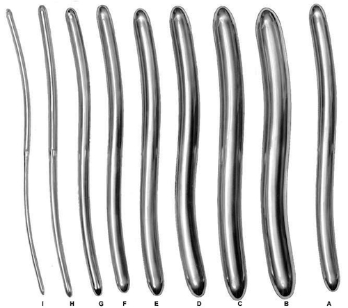
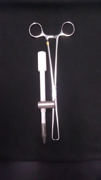
The current procedure for dilating a cervix requires the doctor to use progressively thicker dilators until the desired diameter is reached. This method is very tedious for the surgeon and may put patients at a higher risk for a uterine perforation. To decrease the risk of a uterine perforation, we are going to make a device that, once inserted through the cervical canal, can be controlled by a surgeon to radially dilate the cervix to a desired diameter as indicated on a dial.

Team Picture

Team members from left to right: Michael Martinez, Alex Schmidt, Megan Courtney, Ryan Lane

Images

Female Reproductive Anatomy with specifications.
A set of Hegar Dilators. These are the current devices used to dilate the cervix. Sizes run on a French scale, from a 1 mm size to a 18 mm size in diameter.
The conical cylinder features a threaded portion in which the surgeon would be able to fit this device through a separate part and turn the cone to slowly proceed with dilation.
This separate part, named the phlange, would be attached to two different surgical tools (tenaculums). The surgeon would insert the conical dilator through the hole (which features thread for screw-like movement). This controls the movement to proceed with dilation.
Pigs uterus was used to test our prototype.
Final prototype as received from manufacteurer. Shown is tenaculum, phlange, and cone thread.

Files

Contact Information

Team Members

  • Alexandra Schmidt - Team Leader
  • Ryan Lane - Communicator
  • Megan Courtney - BSAC
  • Michael Martinez - BWIG

Advisor and Client

  • Prof. Randolph Ashton - Advisor
  • Dr. Dan Lebovic - Client
Back to Top