
Engineering a biomimetic Intestine for 3D traction force studies
Project Overview
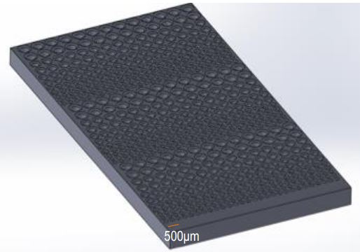
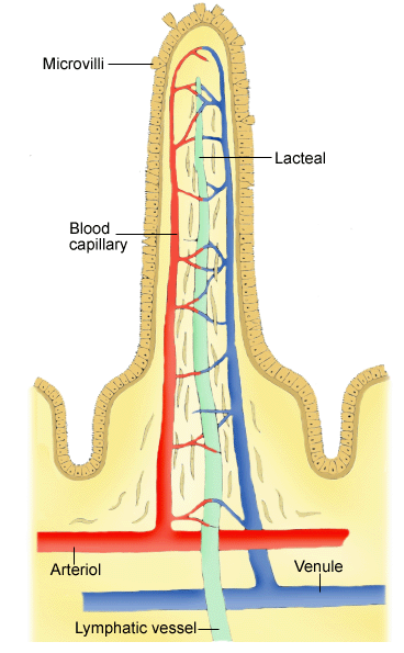
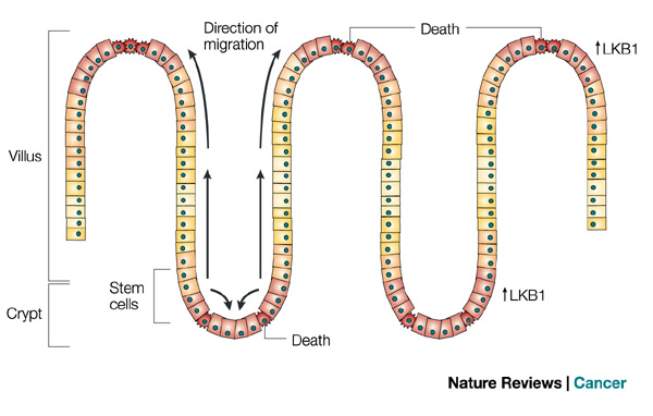
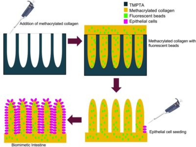
The intestinal epithelium, a series of folds and invaginations into the intestinal lumen, is the primary means of nutrient absorption. These finger-like folds called villi are composed of extracellular matrix (ECM) and serve as a substrate for epithelial cell development. At the crypt or base of each villus, stem cells are continuously being reproduced. As young cells migrate upward toward the tips of the villi, they differentiate into mature epithelial cells. Mature cells are then extruded from the apex to keep this proliferation in check. Our client, Professor Michael Murrell’s goal is to study the mechanical stresses these epithelial cells produce throughout their migration in the hope to determine the mechanism for this collective cell movement. To accomplish this, it is our responsibility to design and manufacture a 3D master mold that is biologically representative of the intestinal villi. From this, we will generate a collagen scaffold to be coated with epithelial cells in order to conduct the traction force study.
Team Picture
Images
Files
- Poster (December 10, 2013)
- Midsemester Oral Report (October 4, 2013)
- Final Report (December 11, 2013)
- PDS (December 11, 2013)
- Midsemester Report (October 9, 2013)
Contact Information
Team Members
- Conor Sullivan - Team Leader
- Susanna Kwok - Communicator
- Kevin Knapp - BSAC
- Shaun Pomerenke - BWIG
- Angela Beltrame - BPAG
Advisor and Client
- Prof. Randolph Ashton - Advisor
- Prof. Michael Murrell - Client