Apparatus for fusion of surface ultrasound and x-ray fluoroscopy images in cardiac procedures
This project has been secured to protect intellectual property.
Login for More InformationProject Overview
Recently, image fusion between 3D cardiac ultrasound (Echo) and X-ray fluoroscopy (XRF) has been proposed to guide the placement of devices such as artificial heart valves. XRF provides high quality real-time imaging of the metal structures in the devices. 3D Echo provides real-time imaging of cardiac soft tissue anatomy and blood flow. In essence, Echo/XRF fusion combines soft tissue imaging from echo with device visualization from XRF, in order to enable proper positioning of the devices.
Most of the current reports demonstrating the concept of Echo/XRF image fusion have focused on the trans-esophageal echo (TEE) probe, which enters the esophagus and performs ultrasound imaging from inside the patient. Registration between ultrasound and XRF images is achieved by detecting the probe orientation within the x-ray images. Unfortunately, TEE is invasive, requires general anesthesia, and poses significant patient risks. On the other hand, surface echocardiography is completely non-invasive. Surface echo is performed with an external probe placed against the chest wall. Despite lower image quality than TEE, surface echo maybe be better suited for routine cardiac interventions, due to its lack of invasiveness and high availability in hospitals.
The fusion of surface Echo and XRF has not yet been explored, despite the fact that surface ultrasound is more common than TEE. The main technical challenge with surface Echo/XRF fusion is that the external probe may not be visible in the XRF field-of-view, or if it is visible, it may not be possible to accurately determine its position and orientation from the XRF image. To solve this problem, the external ultrasound probe must be outfitted with x-ray-visible metal fiducials which are rigidly attached to the probe and which extend into the x-ray image. The detected orientation of these metallic fiducials can be used to encode the position and rotation of the external probe.
Our Mid-Semester Presentation:
https://uwmadison.box.com/s/p61obqdmfeuw72sf2hoagno0a1p72lj2
Images of a proposed design from a BME grad student:
https://dl.dropboxusercontent.com/u/16409236/BME_2015_Proposal_Drawing.png
Paper discussing the merits of similar fiducial designs:
http://scitation.aip.org/content/aapm/journal/medphys/32/10/10.1118/1.2047782
Video Demonstration of TEE:
https://www.youtube.com/watch?v=40iHkbPTcb0
CAD drawing of potential fiducials:
bmedesign.engr.wisc.edu/projects/projectbuilder/info/
Relevant Paper:
http://scitation.aip.org/content/aapm/journal/medphys/32/10/10.1118/1.2047782
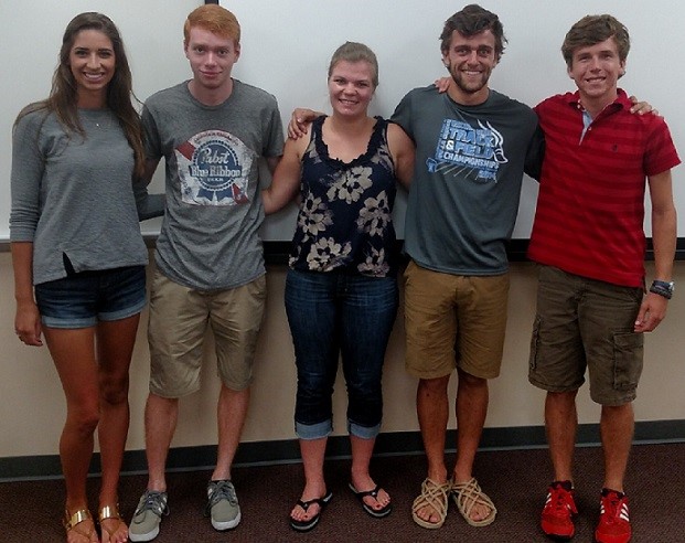
Team Picture

Contact Information
Team Members
- Ashley Hermanns - Team Leader
- Ellen Restyanszki - Communicator
- Alec Onesti - BSAC
- Andrew Duplissis - BWIG
- Nathaniel Bennett - BPAG
Advisor and Client
- Prof. Walter Block - Advisor
- Dr. Amish Raval - Client
