Model for teaching thoracocentesis in dogs
This project has been secured to protect intellectual property.
Login for More InformationProject Overview
Thoracocentesis is the procedure of removing air or fluid from the pleural space (between the lungs and the chest wall). This procedure is done to relieve respiratory distress and to obtain diagnostic samples.
To assist with teaching veterinary students and new graduates, this project seeks to create a realistic model of the canine thorax to simulate the procedure of thoracocentesis. The components that are most important include: thoracic wall (ribs, muscles, skin), space between the lung and thoracic wall, and simulated lung tissue (that might be capable of demonstrating injury if the procedure was not done correctly). The model should be reused by multiple classes.
The general procedure is described in detail in this link: https://cvm.ncsu.edu/wp-content/uploads/2016/11/Therapeutic-thoracocentesis-technique-fenestrated-catheter.pdf
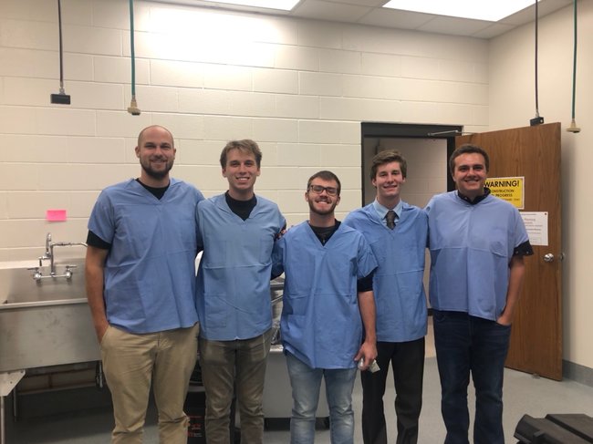
Team Picture

Contact Information
Team Members
- Robert Weishar - Team Leader
- Nicolas Haller - Communicator
- Jacob Reiss - BSAC
- Frank Seipel - BWIG & BPAG
Advisor and Client
- Prof. Tracy Jane Puccinelli - Advisor
- Dr. Julie Walker - Client
Related Projects
- Spring 2019: Model for teaching thoracocentesis in dogs
- Fall 2018: Model for teaching thoracocentesis in dogs
