Flexible microsurgical background
This project has been secured to protect intellectual property.
Login for More InformationProject Overview
Creating an optimized operative field for microsurgery is largely dependent on adequate visualization of vessels or nerves to be
anastamosed. Two crucial aspects of optimized visualization are a high contrast environment and egress of fluid buildup in the
operative field. Currently, a microsurgical "background" device exists, which is a rigid piece of suction tubing fitted to a bright
yellow or blue piece of meshed silicone, allowing for constant suctioning of fluid beneath a highly contrasted background mat.
Unfortunately, this background tubing is overly rigid such that it will consistently shift, move, and flip over, becoming unusable and
requiring constant repositioning.
As such, the surgeons here at UW have created an improvised flexible microsurgical background consisting of a piece of
Xeroform mesh (yellow, semipermeable petroleum infused gauze) wrapped around a pediatric feeding tube, which is affixed to
low suction. This device serves the same function without the drawbacks of an overly rigid device. Its improvised nature, however,
makes it time consuming to repeatedly create.
Our project would propose creating a flexible microsurgical background that could be packaged and reproduced to allow the
advantages of a high contrast background and constant suction without the disadvantages of being overly rigid.
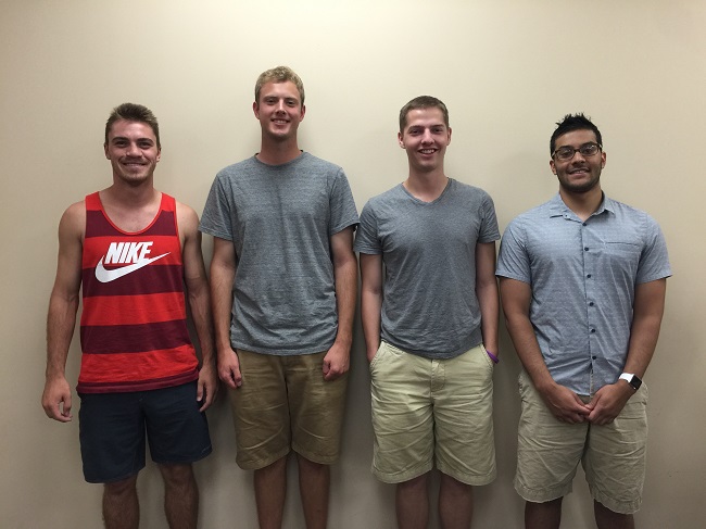
Team Picture

Contact Information
Team Members
- Michael Wolff - Team Leader
- Shakher Sijapati - Communicator
- Ross Barker - BSAC
- Michael Lohr - BWIG & BPAG
Advisor and Client
- Prof. Paul Thompson - Advisor
- Brian Christie - Client
- Prof. Justin Williams - Alternate Contact
Related Projects
- Spring 2016: Flexible Microsurgical Background
- Fall 2015: Flexible microsurgical background
- Spring 2015: Flexible microsurgical background
