Multidimensional imaging-based models for cardiovascular procedural skills training
Our goal is to build a canine heart model for veterinary students to practice balloon valvuloplasty and stent placement for pulmonary stenosis.
Project Overview
Interventional cardiology is a rapidly expanding field in veterinary medicine. Pulmonary valve stenosis occurs when a dog is born with a malformed pulmonary valve, which restricts blood flow from the right heart to the lungs. Balloon valvuloplasty is a palliative procedure in which a balloon-tipped catheter is inserted into the jugular vein to the valve and is then inflated to help reduce the severity of the stenosis. Recently, the UW-Madison School of Veterinary Medicine has experienced a decrease in caseloads of canines with pulmonary valve stenosis, preventing the cardiology residents from being able to practice repairing this disorder. There is a need for a heart model to mimic pulmonary valve stenosis for residents to learn and practice repairing these valves.
This device, a model-based simulation program will be implemented to maintain the cardiologists’ surgical skill set and to aid in cardiology resident training. Simulator training using multidimensional imaging-based models will augment the training already provided in the interventional lab and help protect against the ebb and flow of procedural caseload eroding skills. It also provides a more consistent experience for our residents and provides an objective method of assessing individual progress amongst our trainees.
The goal is to develop a silicone 3D model of canine pulmonary valve stenosis which can be used to learn/practice essential skills like handling of guidewires/catheters, balloon positioning and inflation, and communication between veterinary interventionists. Computed tomography angiography (CTA) of dogs with pulmonary valve stenosis will be used to create the 3D models, which will be secured in place. Lastly, a document camera will project an image of what the user is doing with their hands onto a screen. This provides a more realistic recreation of the interventional surgery, where the surgeon watches a fluoroscopy screen to monitor the movement of the interventional equipment inside the patient.
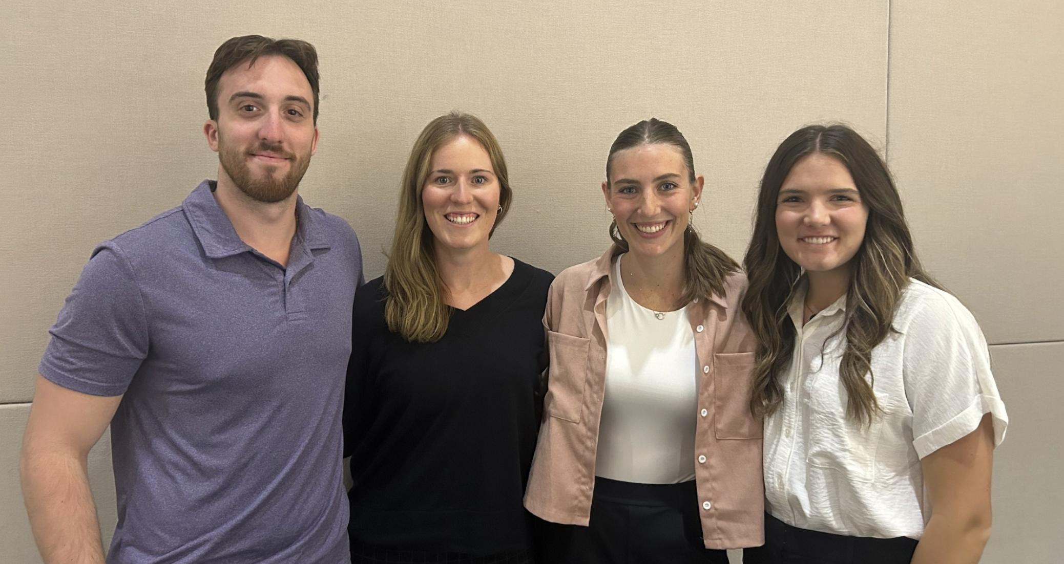
Team Picture

Files
- Final Report (December 8, 2024)
- Final Poster Presentation (December 5, 2024)
- Preliminary Report (October 9, 2024)
- PDS (September 19, 2024)
Contact Information
Team Members
- Hunter Belting - Team Leader
- Anna Balstad - Communicator
- Rebecca Poor - BSAC
- Daisy Lang - BWIG & BPAG
Advisor and Client
- Prof. Tracy Jane Puccinelli - Advisor
- Dr. Sonja Tjostheim - Client
Related Projects
- Spring 2025: Multidimensional imaging-based models for cardiovascular procedural skills training
- Fall 2024: Multidimensional imaging-based models for cardiovascular procedural skills training
