
Radiographic radiomics phantom development
Design and construction of a phantom suitable for radiomics benchmarking in radiographic imaging systems
Project Overview
Radiomics, the extraction and analysis of high-dimensional quantitative texture features from medical images, has shown promise in oncology for prognosis, treatment response assessment, and outcome prediction. However, radiomic features are highly sensitive to image acquisition specifications and hardware specific characteristics. In simpler terms, models developed on one imaging platform cannot be reliably applied to another without proper validation and benchmarking. This benchmarking requires a phantom containing a variety of heterogeneous, tissue-like textures, however commercially available phantoms lack a sufficient range of materials and are not designed for this purpose.
Our client, Dr Jennie Crosby, requires a phantom that incorporates multiple clinically relevant texture patterns, is compatible with imaging protocols for both small anatomy (e.g., head) and large anatomy (e.g., pelvis), and is constructed from materials stable over a multi-year period to support longitudinal radiomic feature evaluation across radiographic imaging systems.
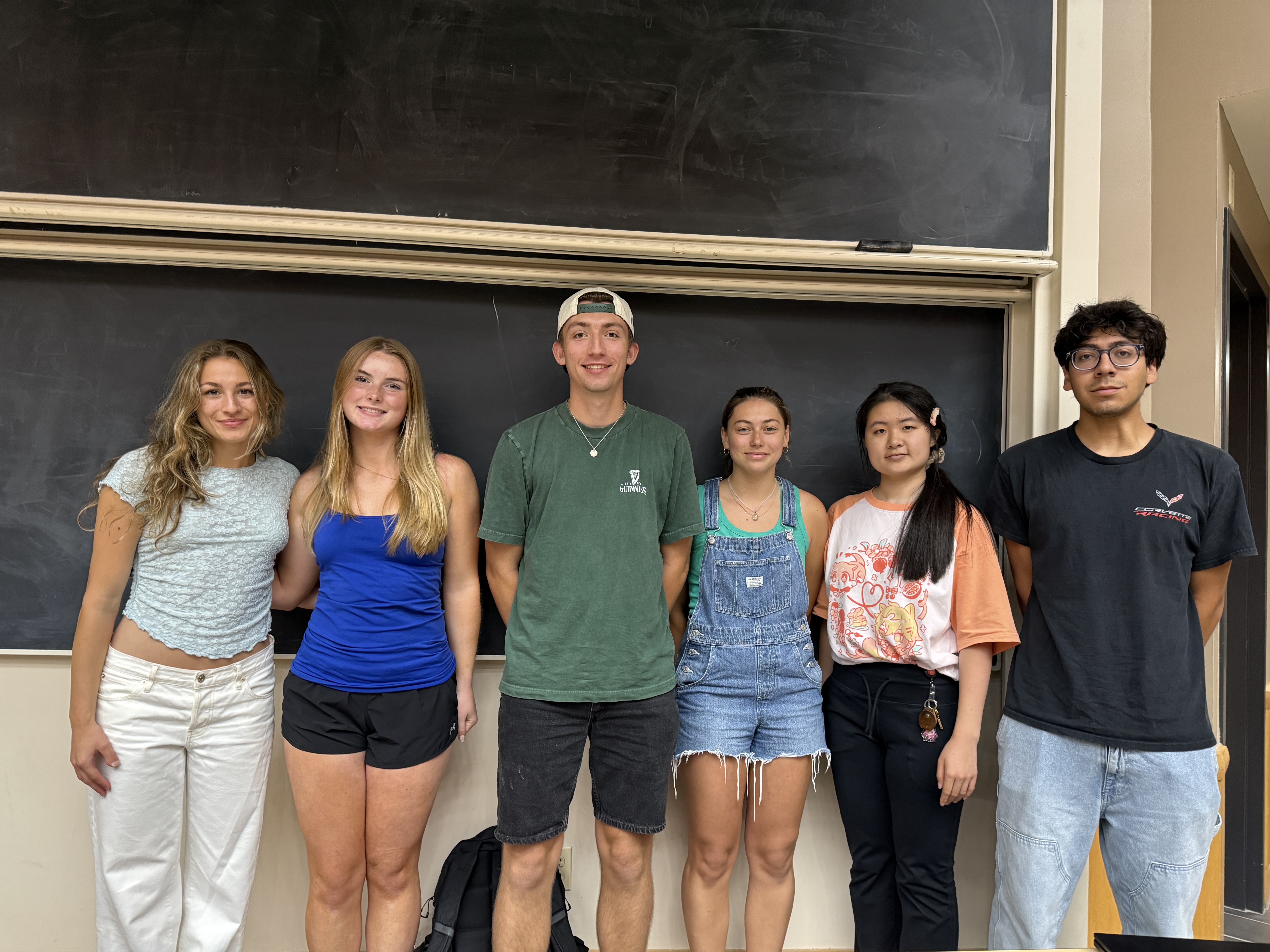
Team Picture
Files
- Preliminary Report (October 7, 2026)
- Preliminary Presentation (October 2, 2026)
- PDS (October 2, 2026)
Progress Reports
- Week 7 (October 22, 2026)
- Week 6 (October 15, 2026)
- Week 5 (October 8, 2026)
- Week 4 (October 1, 2026)
- Week 3 (September 24, 2026)
- Week 2 (September 17, 2026)
- Week 1 (September 10, 2026)
Contact Information
Team Members
- Pierson Trapskin - Team Leader
- Ava Hodgson - Communicator
- Presley Stellflue - BSAC
- Matthew Casas - Co-BWIG
- Sonja Pionek - Co-BWIG
- Allie Ma - BPAG
Advisor and Client
- Prof. Yang Lu - Advisor
- Dr. Jennie Crosby - Client