MRI-Compatible Bioreactor for Cancer Cells

Project Overview

The purpose of staging cancer is to describe the severity and extent of the malignancy. Stage is one of the most important factors that oncologists consider when determining treatment plans and prognoses for their patients. There is a potential to use information about the metabolic state of cancer cells to characterize their stage. One way to follow this metabolism noninvasively is to implement magnetic resonance imaging (MRI) to track hyperpolarized carbon-13 labeled pyruvate as the cells break it down. We aspire to create a bioreactor that will permit a cell scaffold containing malignant cells to be imaged by an MRI scanner. This system will be supplemented with equipment capable of flowing the necessary gases and substances through it for testing.

Team Picture

Team Members (left to right): John, Sarah, Sam, and Jeff

Images

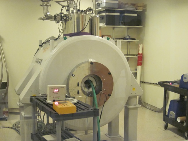
Small-Animal MRI Scanner: The bioreactor cartridge will be placed into the horizontal bore for scanning.
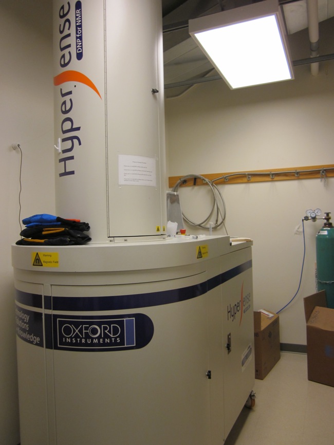
C-13 Hyperpolarizer: This machine hyperpolarizes the C-13 used in the metabolism studies. The tubing that goes into the wall transfers the solution to the MRI room to increase injection speed.
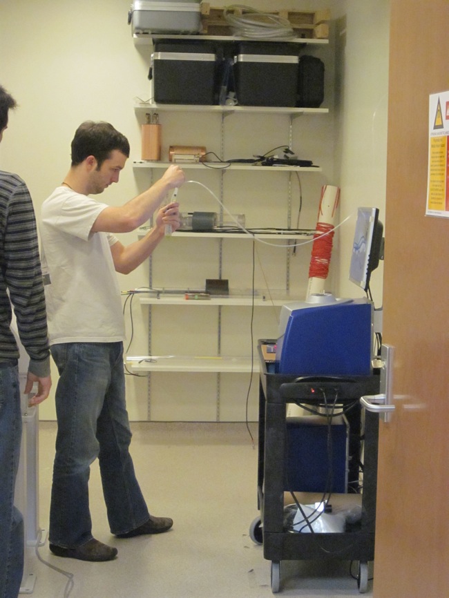
Jeremy Gordon, one of Dr. Fain's graduate students, obtaining the hyperpolarized C-13 solution to be injected into a laboratory mouse for MR scanning and data collection.
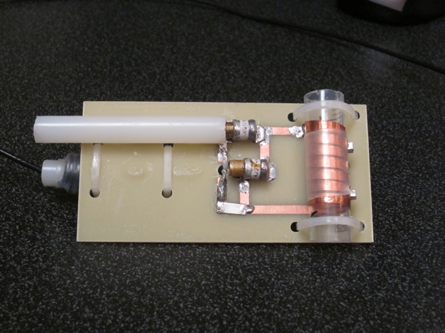
MR Coil: an example of a coil used to collect data with the MR scanner. The bioreactor cartridge will be slid into a copper solenoid, like the one on the right of the image. The more tightly the coil fits around the cartridge, the higher the resolution will be of the resulting scan.
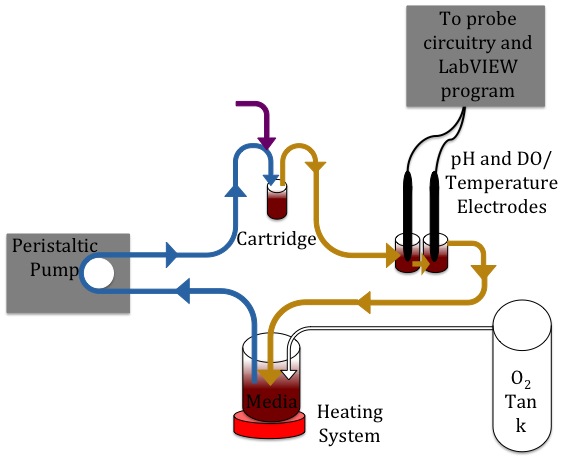
Diagram of Bioreactor System: Components and direction of flow for the integrated system. Blue lines indicate active pumping. The purple arrow represents the injection port. Yellow lines indicate passive flow. The white arrow is for the active pumping of O2 into the system.
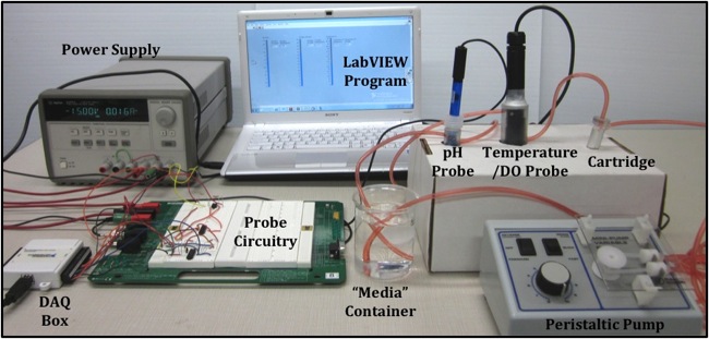
Integrated Bioreactor System: The final prototype for this semester. The peristaltic pump drives the media in a closed loop through the cartridge and sensing containers with the probes. The output of the probes is transmitted and manipulated by the circuitry and then fed into the LabVIEW program for display.

Files

Contact Information

Team Members

  • Jeffrey Hlinka - Team Leader
  • Samantha Paulsen - Communicator
  • John Byce - BSAC
  • Sarah Reichert - BWIG

Advisor and Client

  • Prof. Brenda Ogle - Advisor
  • Sean Fain - Client
Back to Top