
Neonatal thoracic procedural model for thoracentesis and pericardiocentesis, including point of care ultrasound
This project has been secured to protect intellectual property.
Login for More InformationOur goal is to develop a simulator which will allow trainees to, simultaneously, obtain anatomically accurate ultrasound images, and advance a needle into the pleural or pericardial space under ultrasound guidance.
Design Award
- Design Excellence Award Honorable Mention
Project Overview
Thoracentesis (placing a needle into the chest wall through the parietal pleural to evacuate air or fluid) and pericardiocentesis (placing a needle into the pericardial sac to remove air or fluid surrounding the heart) in neonates are traditionally performed by palpation of anatomical features and landmarks and “blindly” inserting a needle into the respective area. Our client, Dr. Adam Bauer, is a pediatrician and neonatology specialist at UW Health. He is interested in creating an anatomically accurate model of the neonatal thoracic cavity, in which medical professionals in-training can use point of care ultrasound (POCUS) to more accurately and reliably visualize needle entry into the appropriate space.
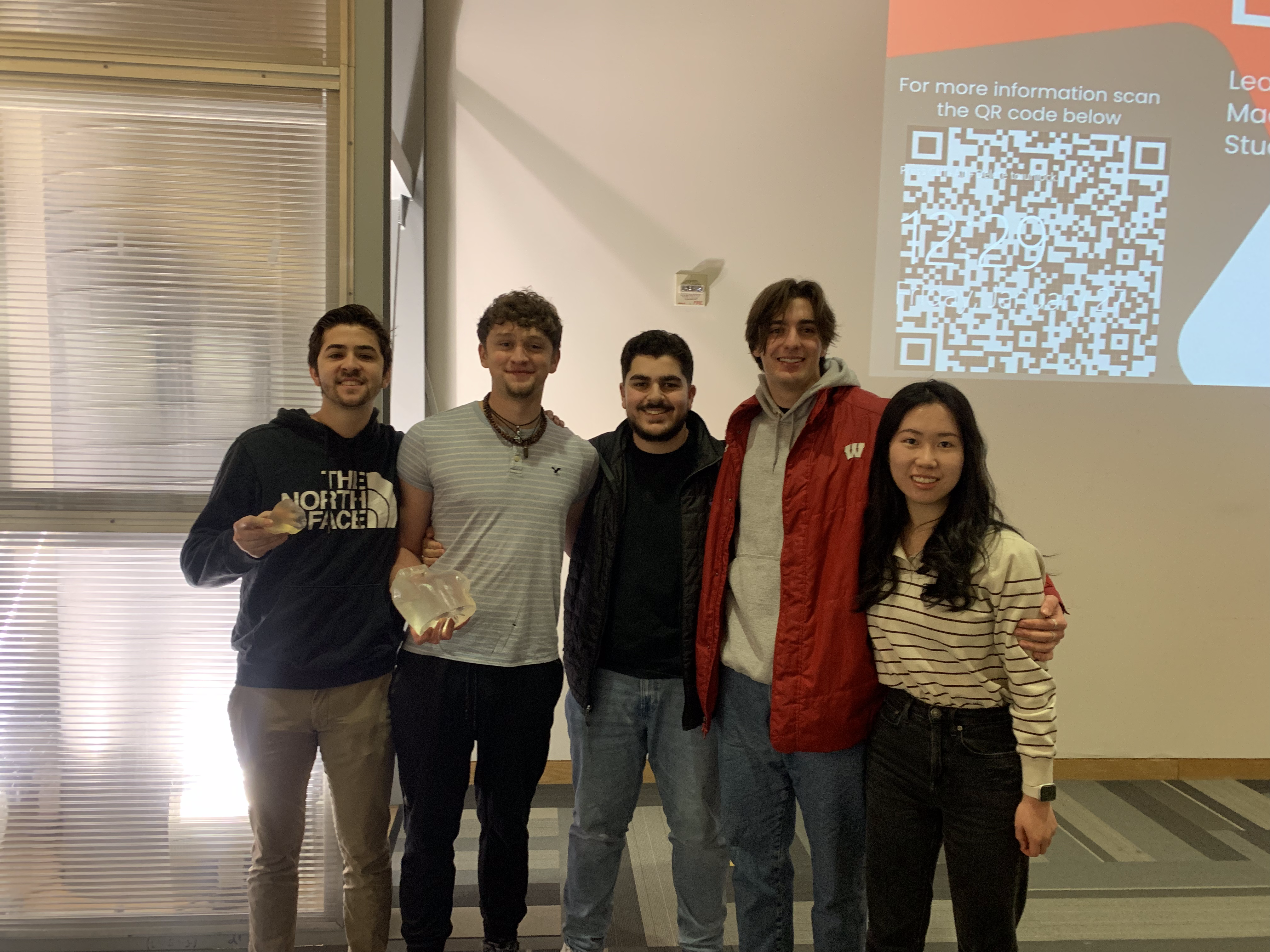
Team Picture
Contact Information
Team Members
- Raad Allawi - Team Leader
- Ethan Runde - Communicator
- Ai Song - BSAC
- Gabriel Piscitelli - BWIG
- Alex Houle - BPAG
- Johanna Ellefson
Advisor and Client
- Dr. Kristyn Masters - Advisor
- Dr. Adam Bauer - Client
Related Projects
- Spring 2023: Neonatal thoracic procedural model for thoracentesis and pericardiocentesis, including point of care ultrasound
- Fall 2022: Neonatal thoracic procedural model for thoracentesis and pericardiocentesis, including point of care ultrasound